The SUIT toolbox is written to support a number of different analysis techniques, such as fMRI group analysis, lesion-symptom mapping, and VBM. All techniques share a number of initial similar steps, including the isolation of the cerebellum from the rest of the brain, normalization to the atlas template, and reslicing of the data into atlas space.
Functions (Matlab version)
- Isolate (suit_isolate_seg)
- Normalize using Dartel (suit_normalize_dartel)
- Normalize with dentate ROI (suit_normalize_dentate)
- Reslice images into SUIT space using dartel (suit_reslice_dartel)
- Reslice images from SUIT space into subject space (suit_reslice_dartel_inv)
- Summarize data in ROIs (suit_ROI_summarize)
Isolate (suit_isolate_seg)
The Isolation algorithm works best on a whole-brain high-resolution (~1 mm isotropic) T1 scan with good gray-white matter contrast. This new version can also combine the information of the T1 scan with other MRI contrast (Channels e.g. T1, T2, PD etc.) to improve the isolation process. The algorithm works best if the T1 image is brought into LPI-orientation, and the origin of the image is set to the anterior commissure.
To isolate the cerebellum simply type suit_isolate_seg in the matlab command window and then
select the appropriate
scan to be analysed using the SPM interface (all steps of analyses
require that SPM is also running). If you want to use different channels, then select all images to be used.
The T1 must be selected first and the following images must have the same dimensions and be coregistered to the
first channel.
To select more than one subject, click in create new subject and then select the corresponding image(s).
Alternatively one can also call suit_isolate_seg({'<name>.nii'}) at the matlab
command window. The input must be a cell array for one subject containing the name of one image per field. The
isolation procedure:
- Segments the brain into tissue-types using John Ashburner's segmentation algorithm
- Crops the full volume, such that it mostly includes the infra-tentorial structures and saves the cropped image as
c_<name>.nii - Uses the tissue-type to calculate the posterior probability of each voxel to belong to cerebellum or brainstem.
- You should check the isolation map visually and if necessary, hand- correct it using a image viewer (e.g. MRICroN, CARET, FSL view)
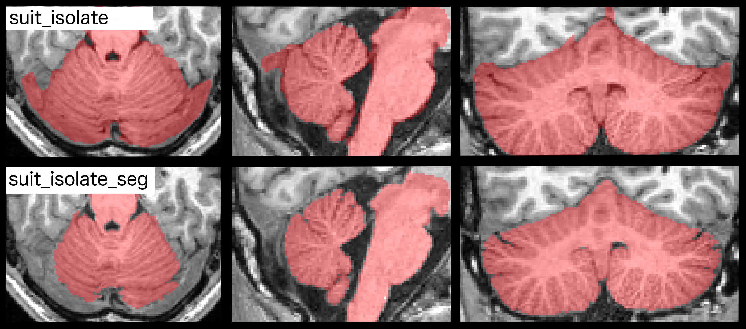
Comparison between cerebellar isolation maps generated by suit_isolate and suit_isolate_seg
Suit_isolate_seg has higher performance that its predecesor. By taking advantage
of the new segmentation implemented in SPM 12, it can better identify the cerebellar boundaries
avoiding misclassification in problematic regions that might result when using the previous version.
Isolation takes about 3 min on a normal desktop PC. Even if
hand correction is necessary, it does not take much more than
5-10 min per individual. For full options of the isolation algorithms
from the command line type: help suit_isolate_seg .
Normalize using Dartel (suit_normalize_dartel)
The next step is to normalize an individual cerebellum into the SUIT atlas template. Human brains differ both in size and shape, the goal of normalization is to deform each person's cerebellum to find the best correspondence with the SUIT template. Compared to normalization to the MNI whole-brain template, the new method greatly improves the alignment of individual fissures, reducing their spatial spread by 60%, and improves the overlap of the deep cerebellar nuclei.
Dartel engine written by John Ashburner uses the tissue
segmentation maps, the white and gray matter segmentation maps produced by suit_isolate_seg
(which uses the segmentation algorithm in SPM12). The non-linear deformation is then found in form of a
flowfield, based on Large Deformation Diffeomorphic Metric Mapping (LDDMM, Michael I. Miller).
To normalize, please specify for each Subject:
- The gray matter probability map (
<name>_seg1.nii) - The white matter probability map (
<name>_seg2.nii) - The (possibly hand-corrected) isolation mask
c_<name>_pcereb_corr.nii
The normalization will produce:
- The affine transformation matrix for the linear part of the normalization:
Affine_<name>.nii - The non-linear flowfield (a 4-D nifti):
u_<name>.nii
No resliced image will be produced at this point. For this, please use suit_reslice_dartel.
Normalize with dentate ROI (suit_normalize_dentate)

Comparison between cerebellar isolation maps generated by suit_isolate and suit_isolate_seg
To investigate activity in the deep cerebellar nuclei, we have developed a version of the SUIT-normalization that uses an ROI from the dentate nucleus to further improve the overlap of the deep cerebellar nuclei (Diedrichsen et al., 2011). As can be seen in the Figure below, this methods leads to exact overlap of the dentate nucleus. This is important for two reasons: First, the dentate nucleus receives input from many functional cerebellar-cortical loops (Dum & Strick, 2003). Thus, when not ensuring good overlap, the activity in these regions will be mixed across participants. Secondly, the raw T2* signal in the dentate nucleus is about 1/2 the size of the BOLD signal of the surrounding gray matter structures (due to the high iron content of the nuclei). Without forcing the dentate nuclei to superimpose during normalization (and without masking the images), cerebellar cortical activation will likely be smoothed into the dentate nucleus.
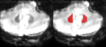
ROI drawing of the dentate "Hull" ROI on the mean EPI image
To normalize a the cerebellum and the dentate nucleus
- Draw an ROI defining the bean shape hypo-intensity on the mean EPI image (or coregistered high-resolution susceptibility-weighted scan)
- Prepare the T1 scan of the cerebellum with
suit_isolateas before - Choose the option "Normalize into SUIT space with dentate ROI", or select the corresponding menu in the GUI.
- Enter the files required for
suit_normalize_dartel, but additionally add the dentate ROI - When reslicing your data into atlas space (using
suit_reslice_dartel), it is useful to use the dentate ROI as a mask. This will prevent any activation to be smoothed into the nucleus - Data in the dentate should not be smoothed before group analysis with more than a 4mm kernel to match the size of the functional units here
- Correction for multiple comparisons over the volume of the dentate nucleus should be applied
Reslice images into SUIT space using dartel (suit_reslice_dartel)
The function suit_reslice_dartel uses the flowfield and affine transformation found by suit_normalize_dartel to bring images into Atlas space.
The Inputs to the function for every subject are:
- Affine transformation matrix: matrix for the linear part of the normalization:
Affine_<name>.nii - Flowfield: The non-linear flowfield (a 4-D nifti):
u_<name>.nii - Images to write: List of images to be resliced. These need to be aligned to the orginal anatomical.
- Cerebellar mask: The (possibly hand-corrected) isolation mask
c_<name>_pcereb_corr.nii
Further options can be specified:
- Modulation: For VBM the intensity should be modulated by the non-linear deformation
- Bounding box: The x,y,z coordinates of the upper and lower corner of the resulting images
- Interpolation: This determines how the image is resampled.
- Filename Prefix: this is the prefix that is prepended to the filename.
For full options of the function see: help suit_reslice_dartel.
Summarize data in cerebellar ROIs (suit_ROI_summarize)
| image | region | regionname | mean | max |
|---|---|---|---|---|
| 1 | 3 | Left_V | 20.1 | 70.7 |
| 1 | 4 | Right_V | 10.3 | 35.4 |
| 1 | 5 | Left_VI | 17.4 | 63.2 |
| 1 | 6 | Vermis_VI | 10.20 | 40.4 |
| ... | ... | ... | ... | ... |
Sometimes a summary of cerebellar data in terms of specific ROIs is very handy. You can use
suit_ROI_summarize to automatically produce such a table. For available Anatomical and functional
atlases, please see our Atlas Collection.
As a first input argument,
you are asked to select the images over which you want the summary. Note that these images need to
be resliced into the same space as the atlas image. The function then finds for each image all voxels within
each of the cerebellar ROIs specified in the atlas file.
The function computes a specific summary statistics (by default
the mean of the data) and generates a text file that can then be used for further analysis (see
right). For full options, see the function help.
For lobular ROIs, you can specify the atlas with
suit_ROI_summarize(images,'atlas','atlas_dir/Diedrichsen2009/atl-Anatom_space-SUIT_dseg.nii');. For backwards compatibility, the function suit_lobuli_summarize is included. However, our recent results (King et al. 2019) show that different lobules do to constitute functional subdivisions of the cerebellum. We
therefore recommend using a good functional parcellation for ROI analyses.
Reslice images from SUIT space into subject space (suit_reslice_dartel_inv)
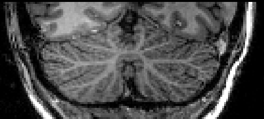
Cropped anatomical image
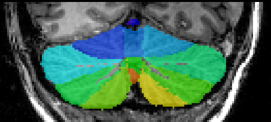
Overlayed with lobular map in anatomical space
The function suit_reslice_dartel_inv takes an image in SUIT space and
reslices it into the space of the individual subject.
This can be especially useful in connection with group atlases (see Atlas Collection) that can be resliced into the original space (see
image).
To reslice back from SUIT space to native space, please specify for each Subject:
- The Affine transformation file created by suit_normalize_dartel
(
Affine_<name>.mat). - The flow field also created by suit_normalize_dartel (
u_a_<name>_seg1.nii). - The reference image for the final geometry (This can be your original or cropped T1 image).
- The image(s) to reslice. If this field is empty the function will automatically reslice the probabilistic atlas into the subject space.