The cerebellum is a functionally highly diverse structure: Different regions have their unique pattern of connectivity with the neocortex, and therefore likely a specialized functional role. To promote accurate anatomical reference for human functional and anatomical imaging studies, we present here a probabilistic atlas of the cerebellar lobules in the space defined by the MNI152 template. The anatomical definitions are based on the fMRI atlas of an individual cerebellum by Schmahmann et al. (2000). To obtain a representative anatomical atlas, we annotated the lobules on T1-weighted MRI scans (1mm isotropic resolution) of 20 individual healthy young participants (10 male, 10 female, average age 23.7 yrs). Using a different set of 23 participants, we also identified the location of the deep cerebellar nuclei. The individual cerebella were then aligned using different commonly used normalization algorithms.
The resultant probabilistic maps allow for the valid assignment of functional activations to specific cerebellar lobules and the nuclei, while providing a quantitative measure of the certainty of such assignments. Furthermore, maximum probability maps derived from these atlases can be used to define regions of interest (ROIs, more on this here). The atlas is included in the newer releases of FSL and the Anatomy toolbox. More versions of the atlases for use with MRIcron or AFNI are also available here.
Please cite the atlas as:
- Diedrichsen J., Balster J.H., Flavell J., Cussans E., Ramnani N. (2009). A probabilistic MR atlas of the human cerebellum. Neuroimage.
- Diedrichsen J., Maderwald S., Küper M., Thürling M., Rabe K., Gizewski ER, Ladd M, Timmann D (2011). Imaging the deep cerebellar nuclei: A probabilistic atlas and normalization procedure. Neuroimage.
The generation of the atlas was supported by a Grant by the National Science foundation (NSF) - BSC 0726685.
Why a probabilistic anatomical atlas?
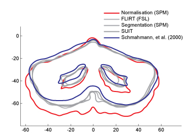
Overlap of the average cerebellar outline after different normalization methods.
Individual brains are often usually normalized to a volume template (such as the MNI template), and activation foci from group analysis assigned to cerebellar lobules using the a standard atlas (Schmahmann et al., 2000). Our analysis shows that such assignments can be incorrect (often by as much as a full lobule) for up to 2/3 of cerebellar voxels for some normalisation methods. There are two reasons for this:
- The Schmahmann atlas is based on a single cerebellum that has its own anatomical peculiarities (for example the right hemisphere extends further down than the left).
- The Colin-cerebellum that forms the basis of the Schmahmann atlas was normalized using an affine registration tool. Other normalisation methods lead on average to a substantially different location of the cerebellum in MNI-space (see image on the left).
Because of this, a representative atlas with good correspondence between the normalization method used for the group analysis and the atlas generation is required. For this reason we provide different versions of the atlas generated with a number of different normalization methods.
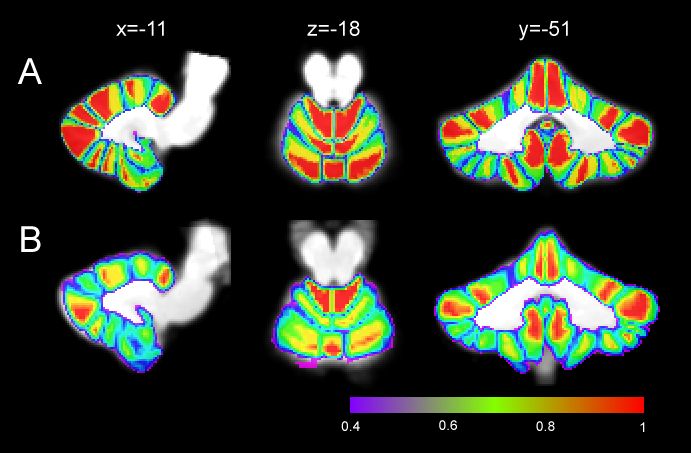
Overlap The overlap in SUIT (A) often reaches 100% whereas the overlap using affine methods (FLIRT, B) is somewhat poorer.
The probabilistic atlas also provides a measure of the certainty with with anatomical assignments can be made. The image shows the proportion of the 20 individuals that overlapped with the same lobule in atlas space. We generated the atlas for:
- Affine alignment after skull stripping in FSL (FLIRT)
- Nonlinear normalization in FSL (FNIRT)
- Segmentation and normalization in SPM5/SPM12 (MNISegment)
- Nonlinear cerebellar-only normalization (SUIT)
- For the old non-linear normalization in SPM2/5
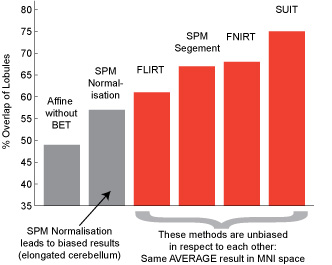
Percentage overlap of individuals with the group atlas after normalization
The left figure shows the percent overlap between different lobules after normalization. Newer non-linear methods (Segmentation in SPM, FNIRT) lead to a good correspondence and to relatively high accuracies. Cerebellum-only normalization with SUIT leads to the best overlap. Thus, for new imaging studies of the cerebellum we would strong recommend one of the newer non-linear methods, or ideally the use of SUIT. For the interpretation of older results (for example for meta-analyses of cerebellar imaging results), we also make the atlas for the SPM nonlinear normalization also available.
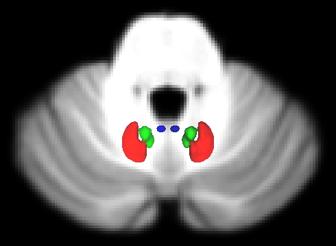
3d reconstruction of dentate (red) , interposed (green), and fastigial (blue) nucleus.
In collaboration with Prof. Timmann from the University of Duisburg-Essen we also imaged the deep cerebellar nuclei in a set of 23 separate subjects at 7T, using susceptibility-weighted imaging. The dentate, emboliform, globose and fastigial nuclei play an important part of the cerebellar circuit, as the relay all output of the cerebellum. In the latest versions of the probabilistic atlas we provide probabilistic ROIs for the dentate, the interposed (emboliform and globose) and fastigial nuclei. Note that given the small size of the cerebellar nuclei, the overlap is relatively poor even after SUIT normalization. For studies of these structures, we therefore recommend spatial normalization using a dentate ROI .
Use the Atlas with FSLeyes
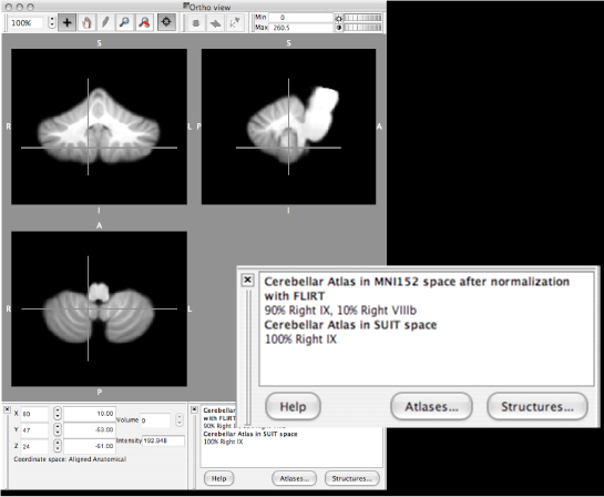
Atlas in altas widget of FSLView.
The atlas can be used with the Atlas Widget in FSLView, part of the FSL package. The atlas for FSLView is available for
- MNI space, using 12-parameter affine alignment to the (new) MNI152-brain-only template (MNIflirt)
- MNI space, using FNIRT, the non-linear normalization to the MNI152 template.
- Spatially unbiased infratentorial and cerebellar template (SUIT)
The FLIRT and FNIRT versions are already included in current distributions of FSL. The lobular assignment is approximately spatially unbiased in these atlases, however the maximal probabilities after SUIT alignment is in general higher. To install the atlas(es):
- download and unpack the zip file
- open a terminal
- navigate to the folder (e.g. cd ~/Desktop/Cerebellum-MNIflirt-FSLView
- run ./install.scp
- the script installs the files in the appropriate folders
The Atlas for MRICroN
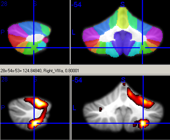
Atlas in MricroN.
The atlas can be used MRIcron, which is an image viewer written by Chris Rorden and which is freely available for Linux, Windows, and Mac OS X.The atlas for MRICroN is available for:
- MNI space, using FLIRT alignment to the MNI152-brain-only template (ATLAS = MNIflirt)
- MNI space, using the standard nonlinear normalization routine in SPM2/5 (ATLAS = MNInorm)
- MNI space, using the segmentation and normalization routine in SPM5/12 (ATLAS = MNIsegment)
- Cerebellar SUIT space (ATLAS = SUIT)
The necessary files (Maximum-probabiliy-map, size of maximum probability, full probability maps, color map (lut) and text files are all included. To use MRICroN to look up probabilities
- open your contrast of interest (bottom panel) on top of the reference image (i.e. SUIT.nii)
- in a separate MRICroN, open reference image (SUIT.nii)
- add Overlap -> Cerebellum-ATLAS.nii.gz. This will show you a color map of the lobules, with the names displayed in the status bar when you click at a certain location in the image. Note this only works with the gz version of the image.
- add Overlap -> Cerebellum-ATLAS-maxprob.nii. Now the corresponding probability of the assignment is also given in the status bar.
- The full probability map is given by Cerebellum-ATLAS-prob.nii with 28 image.
The Atlas for Anatomy Toolbox in SPM
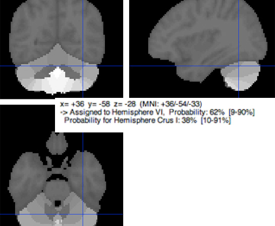
Atlas in Anatomy toolbox (SPM).
The atlas is now included in the Anatomy toolbox, which was developed S. Eickhoff and colleagues. For the generation of the maps, we used the Segementation and Normalisation algorithm in SPM5/8. To make the maps fit with the rest of the toolbox, they were then warped into the space defined by the Colin-brain.
The Atlas for AFNI
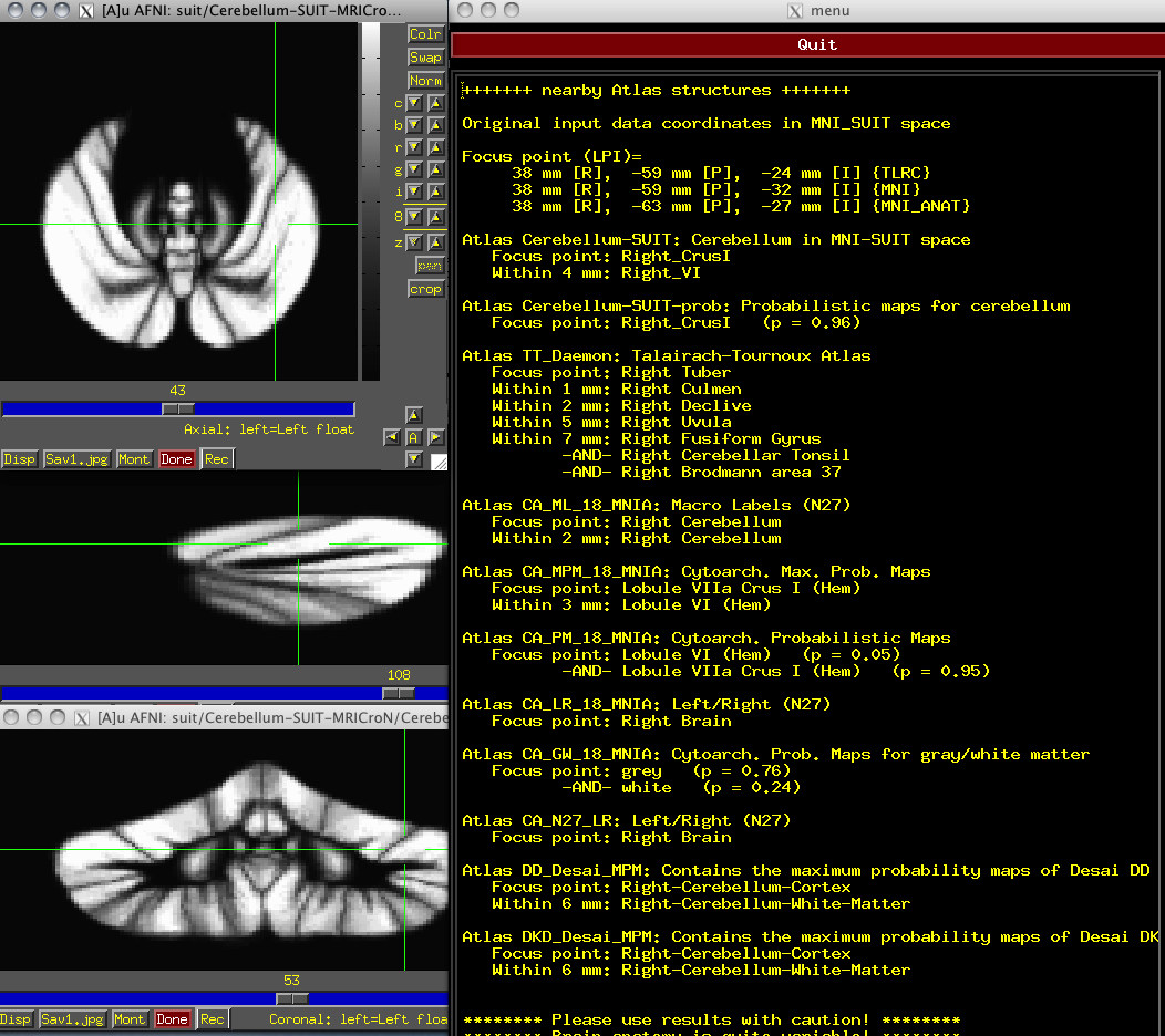
Atlas in AFNI.
Thanks to Daniel Glen, the atlas is now also available for the AFNI whereAmI atlas GUI. Note that any atlas that has a reachable space is queried, so atlases that are in Talairach or MNI_ANAT space are shown there also. The whereAmI output is continuously updated as the user moves the cross-hair in the viewer, so the information is quite interactive. The command line version of whereAmI shows similar information. The command "whereami -show_atlases" gives this relevant output.
To use, please download SUIT for AFNI to a new directory and untar with
tar -xzvf AFNI_SUITCerebellum.tgz
and then add this directory to be searched in AFNI's atlas functions with this:
@AfniEnv -set AFNI_SUPP_ATLAS_DIR directoryname
If you find the atlases and templates useful, please cite using the references included
in whereami -show_atlases
Registration and Download
Note that the SUIT and FNIRT versions of the probabilistic atlas is also included with current release of the SUIT toolbox and in the Atlas Package together with other, functional atlases. For download of a stand-alone probabilistic atlas in different atlas spaces, register below.

The SUIT toolbox distributed under the
Creative Commons Attribution-NonCommercial 3.0 Unported License, meaning that it can
be freely used for non-commercial purposes, as long as proper attribution in form of
acknowledgments and links (for online use) or citations (in publications) are given.
The relevant references are:
Probabilistic atlas for cerebellar lobules
-
Diedrichsen, J., Balsters, J. H., Flavell, J., Cussans, E., & Ramnani, N. (2009).
A probabilistic atlas of the human cerebellum. Neuroimage.46(1), 39-46.

Probabilistic atlas and normalisation for deep cerebellar nuclei
-
Diedrichsen, J., Maderwald, S., Kuper, M., Thurling, M., Rabe, K., Gizewski, E. R., et al. (2011).
Imaging the deep cerebellar nuclei: A probabilistic atlas and normalization procedure.
Neuroimage. 54(3), 1786-1794.
I understand and agree to the above conditions of use.