How to use SUIT for lesion analysis
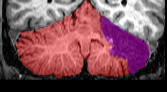
Isolation (Red) and Lesion mask (blue) of an exemplary lesion reconstruction
As a first step, you use suit_isolate to generate
the cerebellar mask (red). How much of the cerebellar lesion
is included in the cerebellar mask is of no consequence - the
lesion will be ignored in the normalization step anyway. Lets
assume the cropped image is call c_patient.nii, and the classification
map patient_pcereb_corr.nii.
Secondly, generate a lesion mask, outlining the
location of the lesion (1 for lesion, 0 otherwise). The mask
can be drawn with MRICro, FSLView, Caret, or any other good
image viewer. Let's say you call this file patient_lesion.nii.
To normalize the cerebellum, we need to tell suit_normalize to ignore the region of the lesion. To do this, the function has to be called from the command line with the option 'lesion_mask':
suit_normalize ('patient.nii', 'mask', 'patient_pcereb_corr.nii','lesion_mask', 'patient_lesion.nii');
Note: The cerebellum mask 'patient_pcereb_corr.nii' should contain the lesioned area also, if this is not the case you can do so using the imcalc function in SPM.
The program then ignores the lesioned area (plus a safety margin of approximately 5mm) in the normalization. Without the safety margin, the nonlinear deformation often "smears" brighter, non-lesioned voxels into the lesioned area, making the lesion on the normalization artificially smaller.
Finally, to bring the lesion mask into the atlas space, we
need to apply the deformation parameters to the lesion mask.
Often, a voxel size of 1 is desired. To do this again call from
the command line:
suit_reslice ('patient_lesion.nii', 'mpatient_snc.mat', 'vox', [1 1 1]);
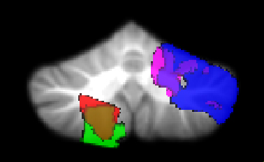
Overlap of lesion from different patients
Using the resliced lesion masks, lesion locations can be compared across different patients. Using imaging programs statistical analysis such as symptom-lesion mapping can be performed based on the normalized lesion masks.